

Robert Gelling
2023-10-03
Excellent treatment and care. Very satisfied!

Paul Hoernel
2023-07-21
A compassionate, supportive, professional staff from the time I set up my initial appointment to my consult, diagnostic scan, my three required and scheduled procedures, treatment ,and post procedure instructions and follow up.Dr.Ninia insures his patients are made to feel comfortable, all options and procedure details fully explained ,and he and his competent support staff help allay any anxiety attached to the procedures from start to finish.Thank you for helping me gain more ‘pep in my step.’
Paul W.Hoernel

margaret donaldson
2023-04-15
From appointment scheduling to treatment .5 stars all around ... this is the place to go !

deb kassay
2022-12-16
So happy I finally had the veins in my legs taken care of. It was easy and painless. Dr. Nina and staff were wonderful to work with and my visits were on time, fast and friendly. Can’t say enough about the results, I am very happy !
Kristi Wilson
2022-12-05
I am so thankful for the staff!!! I needed to take care of my varicose veins for so many Years but I put it off out of fear I finally got into the office and got both legs done a week apart I wish I would have went years ago the staff made me feel so comfortable!!!! I would give them 10 stars if I could!!!

Christina Symelidis
2022-12-05
I think this has been the best experience with a medical provider in my life, and I've had many. Dr. Ninia and his staff were extremely knowledgeable, patient, kind and accommodating. Their work is impeccable. I have a complicated vascular situation and thanks to Dr. Ninia and his staff, I have improved in ways I didn't even expect to when I first came to the office. I can't say enough good things for this wonderful doctor and his practice.
Shannon Urbanski
2022-11-02
Very kind and informative from the get-go! They answered all questions I had and explained exactly how my procedures would go. I’m seriously looking forward to wearing shorts again and getting rid of the leg fatigue!! Highly recommend Dr Ninia & Staff!!

Danielle Cardi
2022-07-07
Dr. Ninia is quick but very thorough. No Wait in the office, which i really appreciate!

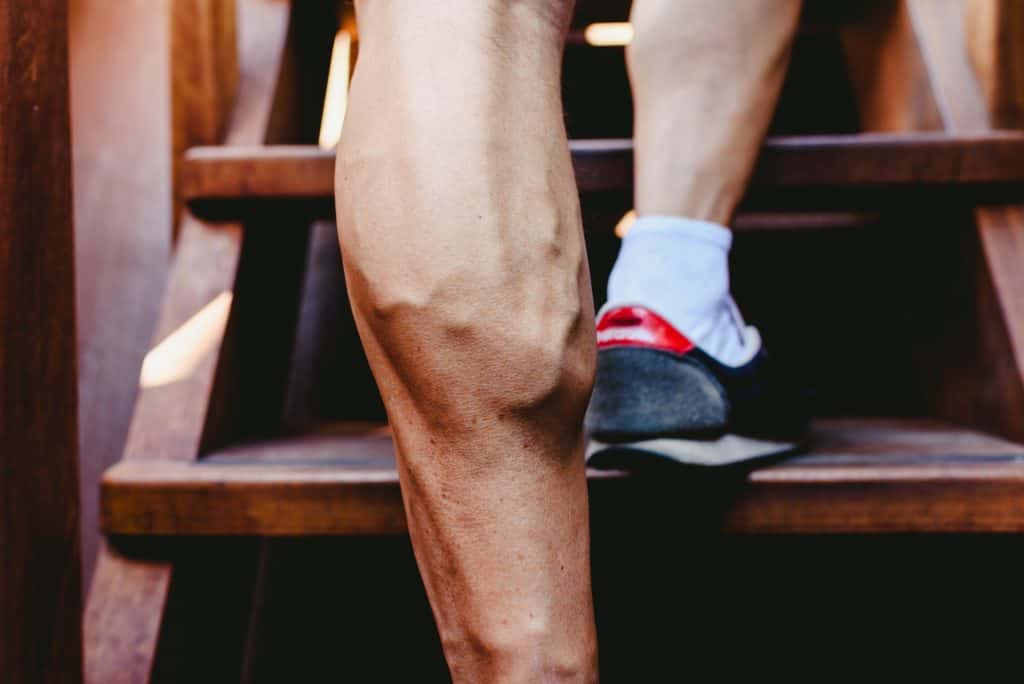
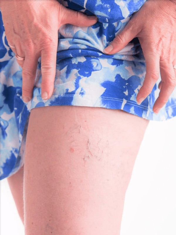
Varicose Vein Removal
Regain confidence with our expert vein removal treatments.

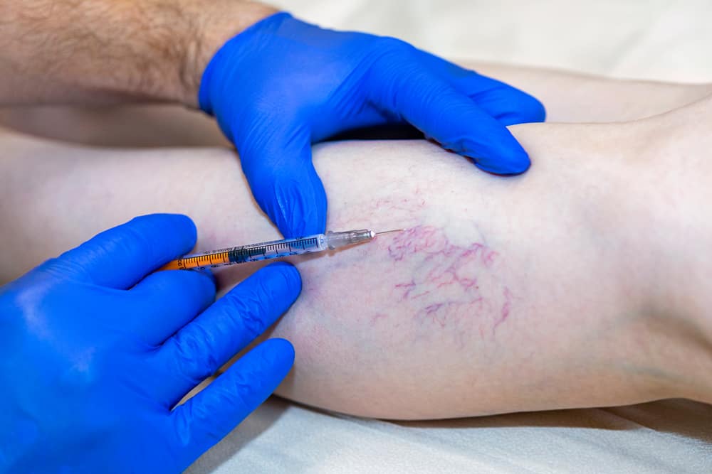
Sclerotherapy
Minimally invasive procedure to treat capillary spider veins.

Laser Ablation
Cutting-edge laser treatments to address larger varicose veins.
VenaSeal
A revolutionary adhesive solution for vein closure without surgery.
Varithena
Advanced foam sclerotherapy ensuring optimal results.

Diagnostic Ultrasound
Safe and painless diagnostic tool to visualize and assess vein health.